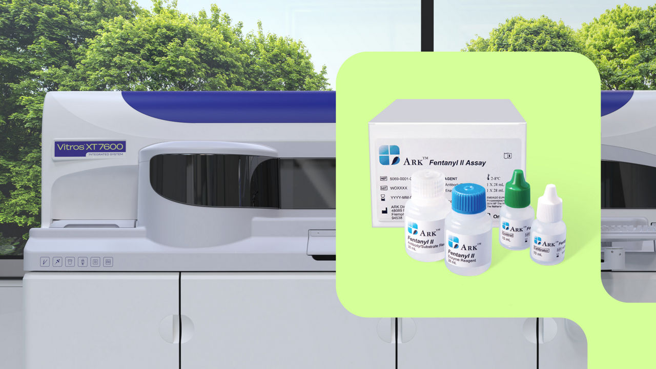
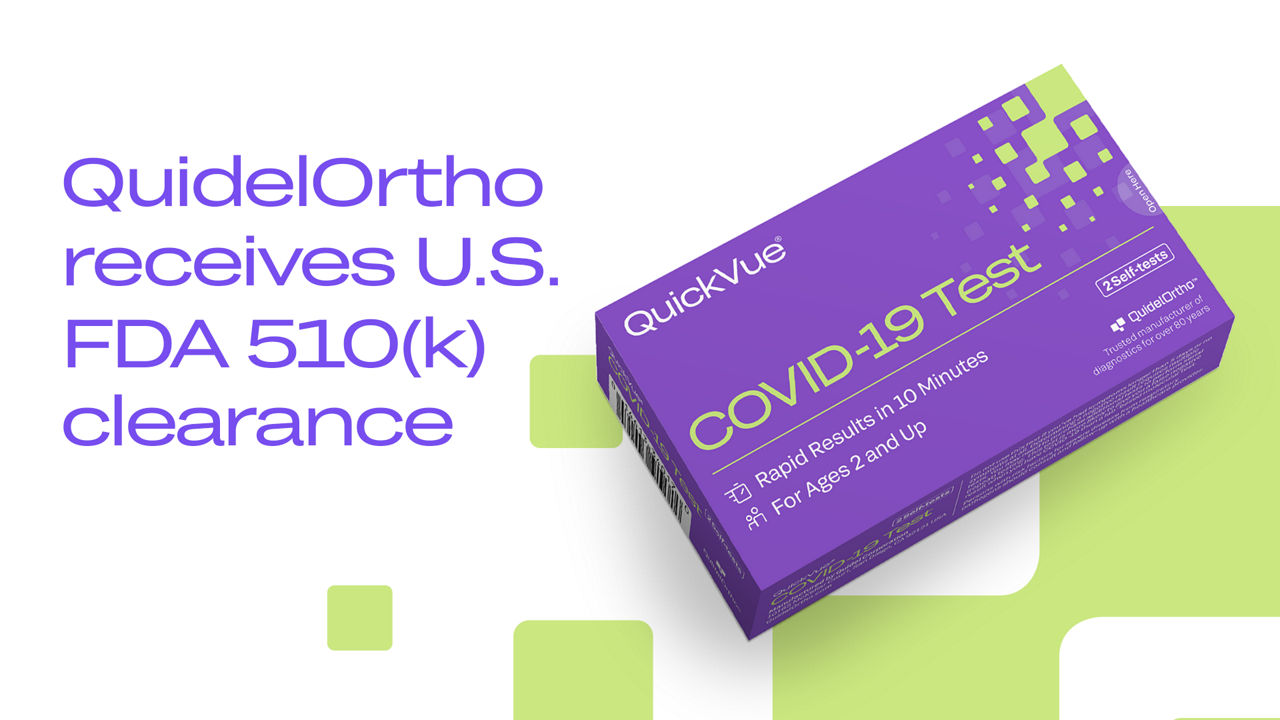
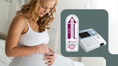
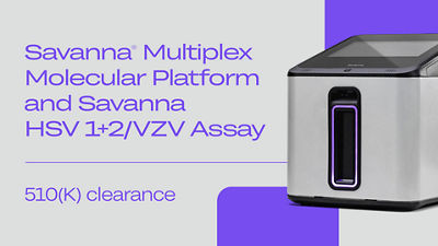
H. pylori
Helicobacter pylori
Helicobacter pylori (H. pylori) is a gram negative rod bacterial pathogen that resides in the mucous layer of the antral and fundal gastric epithelium and occasionally in ectopic gastric tissue in the duodenum or esophagus. H. pylori was first isolated from humans in 1982. The organism is protected from the acidic environment of the stomach by residing in the mucous layer of the stomach and by producing the enzyme urease that breaks down urea into ammonium salts that buffer gastric acidity. H. pylori is highly motile due to its flagella which enables the organism to move within the mucous layer of the stomach.
H. pylori is found in individuals from all parts of the world, but its prevalence is related to geographic region, age, ethnicity and socioeconomic status. Studies suggest that transmission is via the fecal-oral and oral-oral routes. In developing countries, H. pylori colonization is almost universal by age 20. In the United States, prevalence increases with age. Whites are usually not colonized during childhood, but the prevalence of colonization is 50-60% by age 60. Blacks and Hispanics have a higher prevalence of colonization at all ages.
H. pylori is found in 90% of patients with duodenal ulcers and in 50-80% of patients with gastric ulcers. This association with ulcer disease makes H. pylori the most common cause of non-NSAID or aspirin-related peptic ulcer disease. Approximately one in six individuals infected with H. pylori will eventually develop peptic ulcer disease. Other serious complications of infection include atrophic gastritis, gastric adenocarcinoma, and gastric lymphoma. Infected individuals experience long asymptomatic periods with up to 70 percent of individuals having minimal symptoms. Prompt recognition of H. pylori and appropriate intervention and eradication is necessary in order to minimize the potentially serious complications of infection.
Ulcer disease usually presents with gnawing or burning epigastric pain. Other symptoms may include nausea, vomiting, weight loss, bloating, belching and heart burn. Up to 3% of patients with ulcers are asymptomatic.
Testing and identification of H. pylori is important because medications are available that can eradicate this organism. Diagnostic tests for H. pylori fall into two categories: invasive tests that require gastric mucosa samples and noninvasive that do not require mucosa samples. Mucosal samples are usually obtained by endoscopy for biopsy. Tests that do not require a mucosal biopsy include serologic tests, antigen tests using fecal samples, and urea breath tests.
Recommended Reading
Malfertheiner P, Venerito M, Selgrad M. Helicobacter pylori infection: selected aspects in clinical management, 2013, Curr Opin Gastroenterol 29(6):669-75
Karakus, C., and B. A. Salih., Comparison of the lateral flow immunoassays (LFIA) for the diagnosis of Helicobacter pylori infection, 2013, J Immunol Methods 396:8-14.
-
Immunoassay for the rapid qualitative detection of lgG antibodies specific to Helicobacter pylori.
-
The QuickVue TLI H. pylori Stool Antigen Test is a rapid membrane enzyme immunoassay for the qualitative detection of Helicobacter pylori specific...